In this section, we’ll cover undergoing a craniovertebral decompressionA surgical operation which aims to expand the internal dimensions of the craniovertebral junction and, thereby, relieve compression of the neural elements within, and/or to improve the flow of cerebrospinal fluid across the craniovertebral junction.. People who have decided to take up an offer for surgical intervention, for their syringomyeliaA cavity, within the spinal cord, which is filled with cerebrospinal fluid. Syringomyelia cavities come in various “shapes and sizes”, from short, spindle-shaped cavities through to long, tense cavities extending throughout the greater part of the spinal cord. See also hydromyelia. More or Chiari malformationAn anatomical abnormality affecting the lowermost part of brain, where it joins the spinal cord, at the top of the neck. Various sub-types are described – see under their individual names. The term hindbrain hernia is sometimes used to incorporate all forms., may well ask one or more of the questions we propose in this section. These are questions that can also be very reasonably put to their neurosurgeon or a Clinical Nurse Specialist. We have, nevertheless, tried to provide some idea as to what the answers might be.
This varies from one individual to another. The total length of time somebody spends in hospital depends upon several factors, including an individual’s age, what facilities they have at home when they are discharged and whether any complications develop after the surgery. Most people stay in hospital for about a week after the operation, sometimes longer, perhaps as long as two weeks on occasions. This assumes there are no complications. Obviously, the spell in hospital may be a good deal longer if complications develop.
Craniovertebral decompressionA surgical operation which aims to expand the internal dimensions of the craniovertebral junction and, thereby, relieve compression of the neural elements within, and/or to improve the flow of cerebrospinal fluid across the craniovertebral junction. is a major operation for anybody to undergo. The body needs time to recover. It will take at least a month for an individual to feel anywhere near normal again. It is advisable for anybody to assume they will be off work for at least six weeks, perhaps as much as three months. To some extent the period off work is determined by how essential it is for somebody to get back to their employment. Professional people, self-employed and highly motivated individuals tend to get back to work sooner but unless there is a pressing need it is best for somebody to recover adequately. It would not look good for someone appearing not to cope after brain surgery.
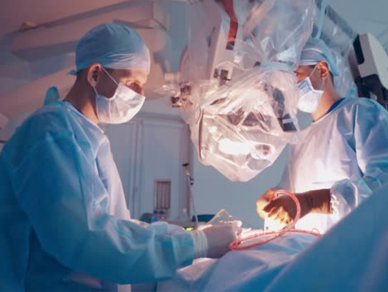
The operating time would be about two hours on average, but the period will vary according to the state of anatomy of an individual case. There is, however, a good deal of preparation to be made, both before and after someone is anaesthetised, before the operation as such begins. After the procedure, when the anaesthetic is reversed, there is a period to be spent in the recovery bay, before a patient returns to the ward. All in all it is likely that you will be in the theatre suite for the best part of a half day.
The wound is about 4″ long, in the midline at the back of the head/upper part of the neck. Quite a few stitches need to be placed deep to the skin, in the muscles and other layers. These will absorb over time. The skin is usually closed with non-absorbable stitches or metallic clips, which need to be removed, usually after about ten days.
Some surgeons choose not to remove hair but most neurosurgeons will take a generous strip of hair in the midline, at the back of the head. Most surgeons feel more comfortable doing this, feeling that the skin can be cleansed and the risk of infection is reduced, although this is not something that can be proven. The head shave also allows for an adequate wound dressing to be applied.
In most cases an outpatient review appointment will be arranged in due course. Ideally this will be six to eight weeks after the surgery but it may be longer, depending upon the pressures on local clinic appointments. Most surgeons would provide emergency access to patients if any problems arose in the meantime.
Craniovertebral decompression aims to correct an internal anatomical abnormality. Many of the symptoms that arise as a result of this condition may improve after surgery. Headache, the most common symptomA symptom is anything of which a patient complains with respect to his or her own body. It may also be obvious to an observer, for example a rash, but it may be entirely subjective, such as a pain somewhere or an emotional feeling., is the one that usually responds well. Even if headaches do not resolve completely, they usually improve significantly. Indeed, craniovertebral decompressionA surgical operation which aims to expand the internal dimensions of the craniovertebral junction and, thereby, relieve compression of the neural elements within, and/or to improve the flow of cerebrospinal fluid across the craniovertebral junction. can be a life transforming operation. Sometimes headaches may return after an interval but they are not usually as severe as they were prior to the surgery. There are a variety of possible causes of recurrent headaches. You would need to discuss matters with your surgeon.
This is a commonly asked question, following intracranial surgery of any type, not just craniovertebral decompressionA surgical operation which aims to expand the internal dimensions of the craniovertebral junction and, thereby, relieve compression of the neural elements within, and/or to improve the flow of cerebrospinal fluid across the craniovertebral junction.. The only restriction is when there is air in the head, which occurs after any intracranial surgery, particularly craniovertebral decompressionA surgical operation which aims to expand the internal dimensions of the craniovertebral junction and, thereby, relieve compression of the neural elements within, and/or to improve the flow of cerebrospinal fluid across the craniovertebral junction.. Theoretically problems could arise if somebody was carried on a high altitude, long haul flight within a short time after such an operation. By a month after surgery any air in the head will be well absorbed. It is unlikely that anybody would choose to take a flight within such a short time after a major brain operation.
In the UK the Driver and Vehicle Licensing Authority place restrictions upon driving following some types of intracranial surgery. The so-called posterior fossa surgery – craniovertebral decompression falls into this category – does not generally require a set period off driving, beyond the requirement for the individual to have recovered in general terms from the surgery. You should not, therefore, plan to drive yourself home from hospital a week after the procedure! It is probably best that you find a “chauffeur” for the first month or so after the operation.
A. There are no strict rules here. Most surgical departments will try to avoid hair washes until the sutures are removed. Even when the stitches do come out the wound is not fully healed by any degree so you should take care when washing your hair. Gentle rinsing rather than vigorous rubbing would be appropriate.
You should consult your General Practitioner. They will almost certainly refer you back to your neurosurgeon who will see you in the clinic and take matters from there.
Even though skin stitches are removed early on, there is a long way to go, both in terms of wound healing and recovery from the surgical trauma. Nature’s healing processes cannot be rushed. It will be at least a month before you feel human again. By three months after the surgery you should have resumed most normal activities. It will be six months, however, before you can push yourself to any degree, e.g. vigorous sporting activity. As regards recovery of lost neurological function this varies. There may be no recovery – the principle aim of the surgery may have been defined as preventing further deterioration, particularly if it is for syringomyeliaA cavity, within the spinal cord, which is filled with cerebrospinal fluid. Syringomyelia cavities come in various “shapes and sizes”, from short, spindle-shaped cavities through to long, tense cavities extending throughout the greater part of the spinal cord. See also hydromyelia. More associated with hind brain hernia. As a general rule of thumb it may take up to two years for recovery of lost neurological function. Beyond that period it is unlikely that further improvement will occur.
A Chiari malformation is a true hernia in the sense that one body part, in this case the tonsils of the cerebellum, protrude through an opening, in this case the foramen magnumThe large, ovoid opening at the base of the skull, measuring about 3 x 3.5cm, through which the spinal cord passes, to continue on as the brain stem. at the base of the skull. A much more common hernia, of course, is one in the abdominal wall or the groin. These can certainly recur if a surgical repair fails. Craniovertebral decompressionA surgical operation which aims to expand the internal dimensions of the craniovertebral junction and, thereby, relieve compression of the neural elements within, and/or to improve the flow of cerebrospinal fluid across the craniovertebral junction. is somewhat different from repair of the abdominal wall. At the craniovertebral junctionThat part of the body where the base of the skull is joined to the top of the spine. Inside these bony enclosures, the craniovertebral junction is also where the brain stem continues downwards as the spinal cord. we are trying to create room for the herniated cerebellar tonsilsThe lower-most part of the cerebellum is made up of a pair of structures, one on each side of the midline, known as the tonsils. These structures have no relationship with the tissue at the back of the throat, which becomes inflamed and sore with a viral infection. It is simply the case that the same Latin term was applied by (presumably) different anatomists, at different times and working in different places, to name these very different body parts. The word tonsilla literally means a stump; Roman ships were moored to “tonsilla” when in port. and to allow CSFCerebrospinal Fluid (CSF), is a clear, colourless liquid that fills and surrounds the brain and the spinal cord and provides a mechanical barrier against shock. Formed primarily in the ventricles of the brain, the cerebrospinal fluid supports the brain and provides lubrication between surrounding bones and the brain and spinal cord. When an individual suffers a head injury, the fluid acts as a cushion, dulling the force by distributing its impact. The fluid helps to maintain pressure within the cranium at a constant level. An increase in the volume of blood or brain tissue results in a corresponding decrease in the fluid. Conversely, if there is a decrease in the volume of matter within the cranium, as occurs in atrophy… More to flow across the craniovertebral junctionThat part of the body where the base of the skull is joined to the top of the spine. Inside these bony enclosures, the craniovertebral junction is also where the brain stem continues downwards as the spinal cord.. We are not trying to repair a weak container. All surgeons will, therefore, try to open up the CSFCerebrospinal Fluid (CSF), is a clear, colourless liquid that fills and surrounds the brain and the spinal cord and provides a mechanical barrier against shock. Formed primarily in the ventricles of the brain, the cerebrospinal fluid supports the brain and provides lubrication between surrounding bones and the brain and spinal cord. When an individual suffers a head injury, the fluid acts as a cushion, dulling the force by distributing its impact. The fluid helps to maintain pressure within the cranium at a constant level. An increase in the volume of blood or brain tissue results in a corresponding decrease in the fluid. Conversely, if there is a decrease in the volume of matter within the cranium, as occurs in atrophy… More channels at the craniovertebral junctionThat part of the body where the base of the skull is joined to the top of the spine. Inside these bony enclosures, the craniovertebral junction is also where the brain stem continues downwards as the spinal cord.. Most will open the membranes that enclose the CSFCerebrospinal Fluid (CSF), is a clear, colourless liquid that fills and surrounds the brain and the spinal cord and provides a mechanical barrier against shock. Formed primarily in the ventricles of the brain, the cerebrospinal fluid supports the brain and provides lubrication between surrounding bones and the brain and spinal cord. When an individual suffers a head injury, the fluid acts as a cushion, dulling the force by distributing its impact. The fluid helps to maintain pressure within the cranium at a constant level. An increase in the volume of blood or brain tissue results in a corresponding decrease in the fluid. Conversely, if there is a decrease in the volume of matter within the cranium, as occurs in atrophy… More. Some will apply heat to the cerebellar tonsilsThe lower-most part of the cerebellum is made up of a pair of structures, one on each side of the midline, known as the tonsils. These structures have no relationship with the tissue at the back of the throat, which becomes inflamed and sore with a viral infection. It is simply the case that the same Latin term was applied by (presumably) different anatomists, at different times and working in different places, to name these very different body parts. The word tonsilla literally means a stump; Roman ships were moored to “tonsilla” when in port., to shrink them in volume. Some surgeons will apply a patch to the membranes that they have opened but others will not. Problems may arise later on if scar tissueIn everyday speech a piece of tissue is a thin sheet of paper, used to wrap a present or, in a slightly different form, to blow the nose, or to be used in the toilet. In biology the word tissue refers to living material made up of cells or groups of cells of similar type or types. We may speak, for example, of nerve tissue or of fatty tissue, of glandular tissue or of connective tissue. Individual organs of the body, such as the brain, the heart, the liver or the kidneys, are made up of several tissue types, almost always including connective tissue. forms at the operation site and causes the surgically created artificial cisterna magna to close down once more, obstructing CSFCerebrospinal Fluid (CSF), is a clear, colourless liquid that fills and surrounds the brain and the spinal cord and provides a mechanical barrier against shock. Formed primarily in the ventricles of the brain, the cerebrospinal fluid supports the brain and provides lubrication between surrounding bones and the brain and spinal cord. When an individual suffers a head injury, the fluid acts as a cushion, dulling the force by distributing its impact. The fluid helps to maintain pressure within the cranium at a constant level. An increase in the volume of blood or brain tissue results in a corresponding decrease in the fluid. Conversely, if there is a decrease in the volume of matter within the cranium, as occurs in atrophy… More movement again. This is not a recurrence of the hind brain hernia as such but it can certainly lead to a recurrence of symptoms. Furthermore, it can be very difficult to treat. Revisional surgery at the craniovertebral junctionThat part of the body where the base of the skull is joined to the top of the spine. Inside these bony enclosures, the craniovertebral junction is also where the brain stem continues downwards as the spinal cord. to post-operative scar tissueIn everyday speech a piece of tissue is a thin sheet of paper, used to wrap a present or, in a slightly different form, to blow the nose, or to be used in the toilet. In biology the word tissue refers to living material made up of cells or groups of cells of similar type or types. We may speak, for example, of nerve tissue or of fatty tissue, of glandular tissue or of connective tissue. Individual organs of the body, such as the brain, the heart, the liver or the kidneys, are made up of several tissue types, almost always including connective tissue., is hazardous, the anatomy is distorted. Verging as against revisional surgery can be likened to the difference between driving on a sunny day and driving through thick fog.
Craniovertebral decompression constitutes major brain surgery. It needs to be carried out in Specialist Neurosurgical Units. These exist in many major cities in the UK but not in all district general hospitals. There are 35 units in the UK, including children’s units.